Anatomy & Physiology
Body Plan
Unlike asteroids, Ophiuroids have a distinct central disc and slender, jointed arms (Ruppert et al. 2004; Hickman et al. 2011; Stohr 2012). Ophiactis savignyi are unlike most other Ophiuroids in that they usually have six arms rather than five. As in all echinoderms, skeletal ossicles are present and can be found in the form of plates (also known as shields) or spines (Ruppert et al. 2004). The arms of O. savignyi consist of a series of sections called articles and each article consists of four shields: an oral shield on the bottom (Figure 1), an aboral shield on the top (Figure 2, 3) and a lateral shield on either side (Figure 2; Ruppert et al. 2004; Stohr 2012). Inside of each article is a vertebra, which is a large ossicle that supports the article, connected by muscle (Figure 2; Ruppert et al. 2004; Hickman et al. 2011). On each article of the arms of O. savignyi, six arm spines are present (three on each side; Figure 1, 2). The underside of the central disc, or oral surface, possesses oral shields that form five moveable jaws around the mouth, each with teeth (Figure 1; Ruppert et al. 2004; Hickman et al. 2011; Stohr 2012). One of these oral shields is modified, forming a madreporite (Figure 1; Ruppert et al. 2004; Stohr 2012). On the aboral side, the central disc is covered with scales and a pair of radial shields are found on either side of each arm (Figure 3; Ruppert et al. 2004). The central disc houses the visceral organs (Hickman et al. 2011).
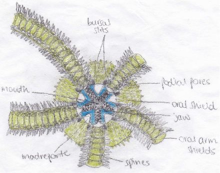
Figure 1: Oral surface of an ophiuroid, adapted from Hickman et al. 2011.
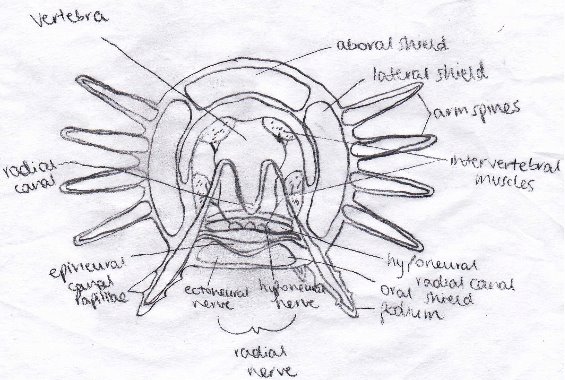
Figure 2: Cross section through the arm of an ophiuroid, adapted from Ruppert et al. 2004.
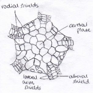
Figure 3: Aboral surface of an ophiuroid, adapted from Ruppert et al. 2004.
The nervous system of an Ophiuroid consists of radial nerves and a nerve ring (Ruppert et al. 2004). This is similar to asteroids, however the nerve ring and the ectoneural cord of the radial nerves have been internalised in the epithelium of the epineural canal (Ruppert et al. 2004). The epineural canal is formed from the enclosed remnant of the ambulacral groove, seen in asteroids, and it lies on the oral side of the radial nerve (Ruppert et al. 2004). Ophiuroids do not have specialised sensory organs, as the sensory system is composed of individual sensory cells (Ruppert et al. 2004).
Digestive System
Ophiuroids have a blind gut that resembles a sac and it is found in the central disc (Hickman et al. 2011). The mouth is in the middle of the oral side of the central disc and is surrounded by five jaws (Figure 1; Ruppert et al. 2004). The mouth is joined to the stomach via a short, yet thick, oesophagus (Ruppert et al. 2004). The stomach is folded, forming ten pouches and it occupies almost the entire interior of the central disc (Ruppert et al. 2004). There is no intestine or anus; therefore any material that cannot be ingested is removed via the mouth (Hickman et al. 2011).
Water Vascular System
As in all echinoderms, Ophiuroids have a water vascular system, which is a coelomic compartment. The water vascular system consists of canals, tube feet, or podia, and dermal ossicles (Figure 2; Hickman et al. 2011). Ophiuroids have a madreporite on the oral surface, which can open to allow water in (Figure 1; Ruppert et al. 2004). The madreporite leads to a stone canal which in turn leads to a ring canal, found on the aboral side of the jaws (Ruppert et al. 2004). The ring canal leads to radial and lateral canals in the arms, with each article of the arms possessing two tube feet, or podia, and a pair of lateral canals (Ruppert et al. 2004). Each of the podia have papillae which are adhesive (Ruppert et al. 2004). There is a valve between the lateral canal and the podia and pressure from fluid protracts the podia (Ruppert et al. 2004). The podia can move by contracting and pumping water through the water vascular system (Ruppert et al. 2004).
|